
Advanced imaging techniques hospital teams use to customize your therapy
Isotope Innovations >> Health>> Advanced imaging techniques hospital teams use to customize your therapy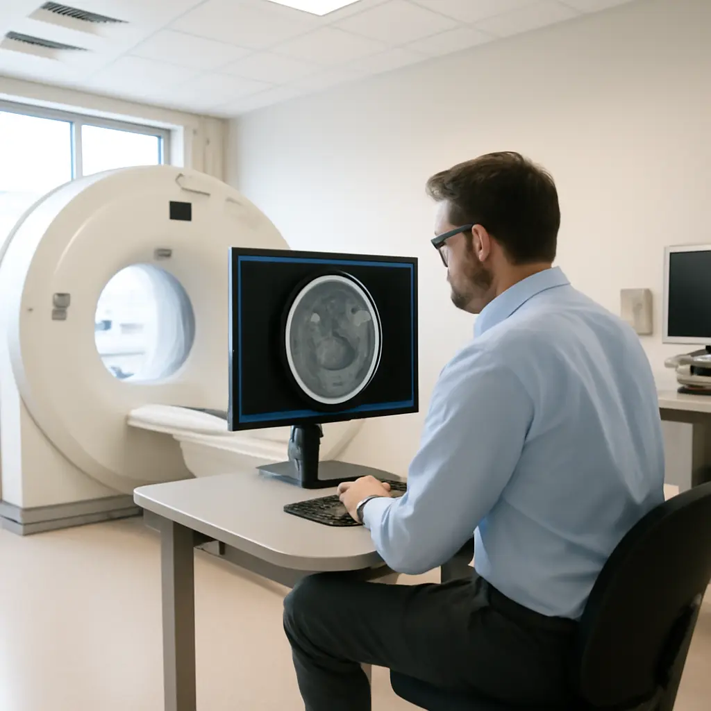
Advanced imaging techniques hospital teams use to customize your therapy
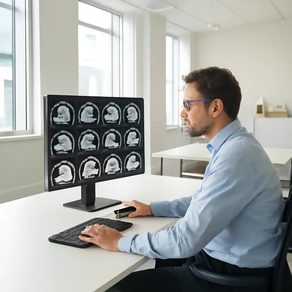
Advanced medical imaging combines technologies such as MRI, CT, PET, ultrasound, and image-guided procedures to show clinicians what is happening inside the body and help them tailor diagnosis and treatment. Rather than relying on a single scan, hospital teams may combine images with laboratory results, pathology, and a patient’s symptoms to select a therapy, plan a procedure, or monitor response. The National Cancer Institute notes that imaging is used to find disease, assess its extent, plan treatment, and evaluate how well treatment is working. These capabilities matter at scale: the International Atomic Energy Agency estimates that about 4.2 billion diagnostic radiology examinations are performed worldwide each year, although access and use vary substantially among countries. The sections below explain how advanced imaging personalizes care, describe major techniques and their limits, and show how teams use imaging in practice.
How advanced imaging customizes therapy
Personalized imaging is the selection and interpretation of medical images to answer a specific clinical question for an individual patient. It is not a single machine or a guarantee that treatment will be more effective. The Radiological Society of North America describes medical imaging as a way to visualize internal structures and help diagnose, guide treatment, and monitor disease. In practice, clinicians combine the images with other evidence before making decisions.
The value of imaging depends on whether its findings can change a decision. A scan may identify the location and size of a tumor, show whether a blood vessel is blocked, distinguish active inflammation from scar tissue, or reveal anatomy that affects surgical access. Radiologists, oncologists, surgeons, nuclear medicine physicians, pathologists, and other specialists may review the results together, especially when a treatment decision is complex.
From diagnostic image to treatment decision
Diagnostic imaging creates information about anatomy, tissue characteristics, or function. CT and MRI commonly provide detailed anatomy; PET and some other nuclear medicine studies show biological activity; ultrasound provides real-time imaging without ionizing radiation. The appropriate test depends on the patient’s symptoms, suspected condition, prior results, and the clinical question. The American College of Radiology’s Appropriateness Criteria help clinicians choose imaging suited to particular scenarios.
Image findings can help teams determine whether to biopsy, operate, use medication, deliver radiation, or observe a condition. They can also help avoid a procedure when imaging and other evidence make it unnecessary. However, no scan independently establishes every diagnosis: findings must be interpreted in context, and some abnormalities require confirmation by biopsy or follow-up.
Personalization has limits
An image is a measurement, not a complete account of a person’s health. Image quality, the scanner protocol, motion, contrast agents, and interpretation can affect what is visible. Incidental findings—unexpected abnormalities unrelated to the original reason for the scan—may lead to additional testing, while false-positive or false-negative results can also occur. Teams weigh these possibilities against the likely benefit of answering the clinical question.
Radiation exposure is another consideration for CT, X-ray, and nuclear medicine examinations. The U.S. Food and Drug Administration advises that imaging using ionizing radiation should be justified and optimized, particularly for children, who are more sensitive to radiation than adults. MRI and ultrasound do not use ionizing radiation, but each has other considerations, including scan duration, availability, implants, patient tolerance, or the need for a contrast agent.
Advanced imaging techniques used to tailor care
Advanced imaging includes more than higher-resolution pictures. Techniques may measure blood flow, tissue diffusion, metabolic activity, electrical or mechanical properties, or the movement of contrast through an organ. These are related approaches, but they answer different questions; a result useful for one disease may not be validated or appropriate for another.
MRI: detailed soft-tissue and functional information
Magnetic resonance imaging uses magnetic fields and radio waves to create images without ionizing radiation. Different MRI sequences emphasize different tissue properties. Diffusion-weighted imaging, for example, measures the movement of water molecules and can help characterize some tumors, strokes, and infections. Functional MRI tracks changes associated with blood oxygenation and is used chiefly to map brain activity, including for selected pre-surgical assessments.
MRI can be useful when clinicians need detailed views of the brain, spinal cord, joints, pelvic organs, or soft-tissue tumors. In cancer care, MRI may help define local tumor extent and provide information for surgical or radiation planning. The National Cancer Institute emphasizes that imaging choices depend on the cancer and the clinical purpose; MRI is not automatically superior to CT or other modalities for every patient.
CT and 3D reconstruction: anatomy for procedures and planning
Computed tomography uses X-rays to produce cross-sectional images, which software can reconstruct into three-dimensional views. CT is often selected when clinicians need rapid assessment of trauma, lung disease, complex bone anatomy, or the location of a lesion relative to nearby structures. These reconstructions can support surgical planning, procedure navigation, and radiation-treatment design.
CT angiography adds contrast material to show blood vessels, while cardiac CT can assess coronary anatomy in appropriate patients. The American College of Radiology cautions through its appropriateness guidance that the best test depends on the clinical scenario. Because CT uses ionizing radiation, teams consider whether the expected diagnostic benefit justifies the exposure and use protocols designed to keep dose as low as reasonably achievable.
PET and SPECT: mapping biological activity
Positron emission tomography (PET) and single-photon emission computed tomography (SPECT) are nuclear medicine techniques. They use radiotracers that emit detectable signals, allowing clinicians to assess selected biological processes rather than anatomy alone. PET/CT combines metabolic or molecular information from PET with structural detail from CT; PET/MRI pairs PET with MRI in settings where the combination is clinically useful.
In oncology, fluorodeoxyglucose PET can reveal areas with increased glucose use, helping assess some cancers, stage disease, or evaluate response. The finding is not cancer-specific: infection and inflammation may also show increased uptake. The Society of Nuclear Medicine and Molecular Imaging therefore stresses that nuclear medicine results must be interpreted alongside clinical history and other tests. Radiotracers and scan protocols are selected for the question being asked.
Ultrasound and elastography: real-time, radiation-free assessment
Ultrasound uses high-frequency sound waves to create real-time images and does not use ionizing radiation. It can guide needle biopsies, drain fluid collections, assess blood flow with Doppler, and examine organs during movement. Because it can be performed at the bedside, it is valuable when a patient is too unwell to travel or when clinicians need immediate procedural guidance.
Elastography is an ultrasound or MRI-based method that estimates tissue stiffness. It is used in selected settings, including assessment of liver fibrosis, and may help clinicians evaluate disease without immediately resorting to biopsy. Results depend on technique and patient factors, so elastography complements rather than universally replaces clinical evaluation or tissue sampling.
Image-guided treatment and monitoring
Imaging can guide care during a procedure as well as inform decisions beforehand. Interventional radiologists and other specialists use live or repeated images to place instruments, target lesions, deliver treatments, and verify results. This connects diagnostic imaging with image-guided therapy, where the image becomes part of the treatment workflow.
Image-guided biopsy and minimally invasive procedures
In image-guided biopsy, ultrasound, CT, MRI, or another modality helps direct a needle to a specific target. The aim is to obtain representative tissue while avoiding nearby structures. Imaging also supports procedures such as drainage, catheter placement, tumor ablation, and embolization. The Society of Interventional Radiology describes image-guided procedures as minimally invasive alternatives or complements to some open surgical approaches, although suitability depends on the condition and patient.
For example, a radiologist may use ultrasound to guide a breast biopsy or CT to reach a lung lesion. The sample then goes to pathology, whose findings may determine whether a patient needs surgery, systemic medication, radiation, or surveillance. Imaging helps target the sample; pathology and the wider clinical assessment help establish what the tissue means.
Radiation therapy planning and delivery
Radiation oncology uses imaging to define a treatment target and nearby organs that need protection. CT is commonly used for treatment planning, while MRI or PET may be registered with the planning scan to add complementary information. During treatment, image-guided radiotherapy can check patient position and anatomy, helping the team account for movement or changes in the treatment area.
The International Atomic Energy Agency describes image-guided radiotherapy as a means of improving localization during radiation treatment. Imaging does not eliminate uncertainty: tumors and organs can move or change, and the clinical team must choose appropriate margins, monitoring, and dose constraints. The treatment plan is individualized according to disease, anatomy, treatment goals, and the patient’s overall condition.
Response assessment and adaptive care
Follow-up imaging can show whether a lesion has changed in size, whether disease has spread, or whether treatment effects are present. In some cancers, metabolic changes on PET may appear before a clear size change on CT. In other situations, scar tissue, inflammation, or treatment-related changes can make response difficult to interpret. Standardized criteria, such as RECIST for certain solid tumors, help clinicians assess changes consistently, but the appropriate method varies by disease and therapy.
In adaptive radiation therapy, clinicians may update a treatment plan when repeat imaging shows meaningful changes in anatomy or tumor position. More broadly, serial imaging can prompt a team to continue, adjust, or reconsider therapy. A scan should be ordered when its likely result could inform care, balancing clinical value with cost, access, radiation exposure where relevant, and the possibility of ambiguous findings.
How hospital teams turn images into individualized care
The pathway from scan to treatment generally involves a sequence of decisions rather than a single technology. A clinician identifies the question, selects an appropriate examination, and considers patient-specific factors such as kidney function, pregnancy, implanted devices, prior imaging, and ability to remain still. A radiologist or other qualified imaging specialist interprets the study and communicates findings to the treating team.
- Define the clinical question and whether imaging is likely to change management.
- Select the modality and protocol that best answer that question while considering risks and alternatives.
- Interpret the images alongside symptoms, examination findings, laboratory tests, and pathology where available.
- Discuss complex cases across specialties and agree on a treatment or follow-up plan.
- Use later imaging selectively to assess response, complications, or changes that could alter care.
A common real-world example is cancer treatment planning. A patient may undergo CT to assess disease extent, MRI to clarify local anatomy, and PET/CT if a metabolic assessment is appropriate. A multidisciplinary team may then combine those findings with biopsy results and the patient’s preferences to plan surgery, radiation, medication, or a sequence of treatments. The modalities are complementary, not interchangeable, and not every patient needs every scan.
Hospitals also use quality controls to support reliable results, including equipment checks, standardized protocols, trained staff, peer review, and comparison with prior examinations. Access to advanced imaging and specialist interpretation is uneven, and delays can affect when decisions are made. The World Health Organization and International Atomic Energy Agency have both highlighted the need for safe, appropriate, and equitable access to diagnostic imaging as part of health-system capacity.
Conclusion: imaging as a tool for more informed treatment
Advanced imaging customizes therapy by connecting detailed anatomy and biological information to diagnosis, procedure guidance, treatment planning, and follow-up. MRI, CT, PET/SPECT, ultrasound, and image-guided interventions each contribute different evidence, while radiation planning and response assessment show how imaging can influence care over time. The central principle is to choose the right examination for a clear clinical question and interpret it with the rest of the patient’s medical evidence.
Patients can ask their care team what question a scan is intended to answer, whether alternatives exist, how the result could affect treatment, and whether previous images can be reused. For further reading, consult the National Cancer Institute’s imaging information, the American College of Radiology’s Appropriateness Criteria, and the U.S. Food and Drug Administration’s guidance on medical imaging. These resources can help patients take part in informed discussions without treating any scan as a substitute for personalized medical advice.
Sources: International Atomic Energy Agency, Diagnostic Radiology, https://www.iaea.org/topics/diagnostic-radiology; National Cancer Institute, Medical Imaging, https://www.cancer.gov/about-cancer/diagnosis-staging/diagnosis/medical-imaging-fact-sheet; Radiological Society of North America, RadiologyInfo.org, https://www.radiologyinfo.org/; American College of Radiology, ACR Appropriateness Criteria, https://www.acr.org/Clinical-Resources/ACR-Appropriateness-Criteria; U.S. Food and Drug Administration, Medical X-ray Imaging, https://www.fda.gov/radiation-emitting-products/medical-imaging/medical-x-ray-imaging; Society of Nuclear Medicine and Molecular Imaging, Patient Resources, https://snmmi.org/Patients; Society of Interventional Radiology, Patients, https://www.sirweb.org/patient-center/; International Atomic Energy Agency, Image Guided Radiotherapy, https://www.iaea.org/topics/radiotherapy/image-guided-radiotherapy; Eisenhauer et al., New response evaluation criteria in solid tumours: Revised RECIST guideline (version 1.1), European Journal of Cancer, 2009, https://doi.org/10.1016/j.ejca.2008.10.026; World Health Organization, Medical devices: Diagnostic imaging, https://www.who.int/health-topics/medical-devices
Related Post

Scanning Too Late Costs More Than Money, Ask Any Oncologist
Cancer screening looks for certain cancers in people who do not have symptoms; diagnostic testing…